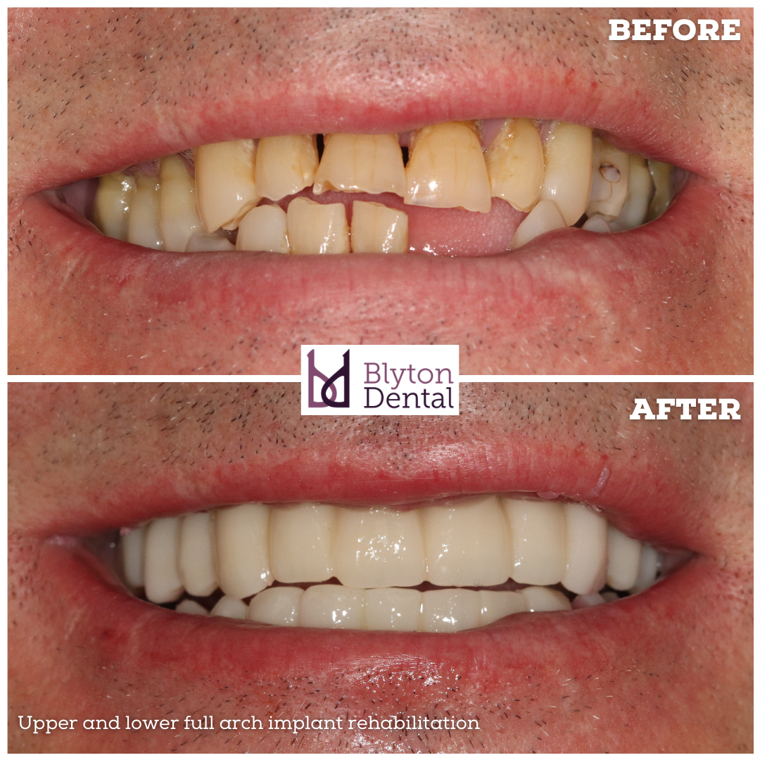
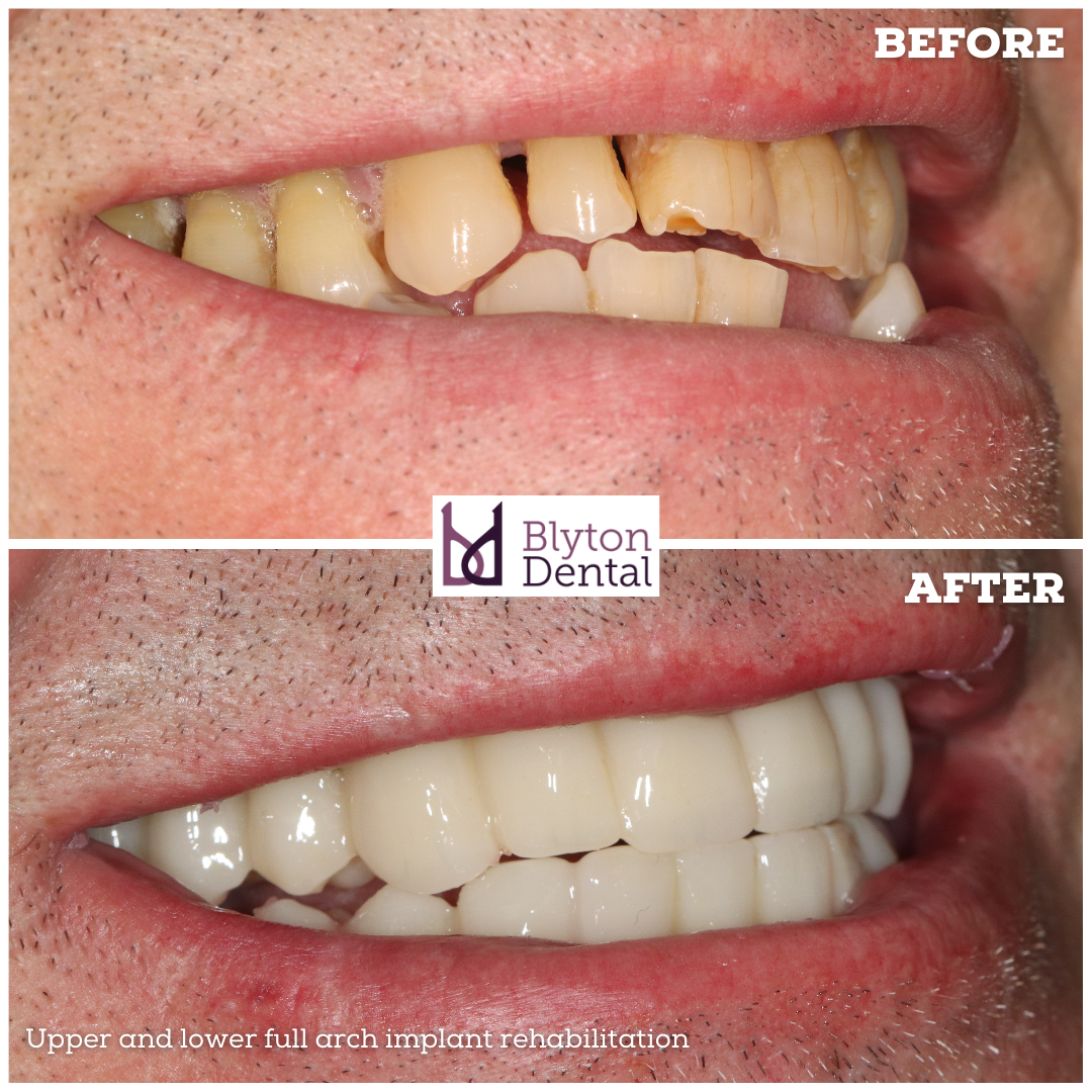
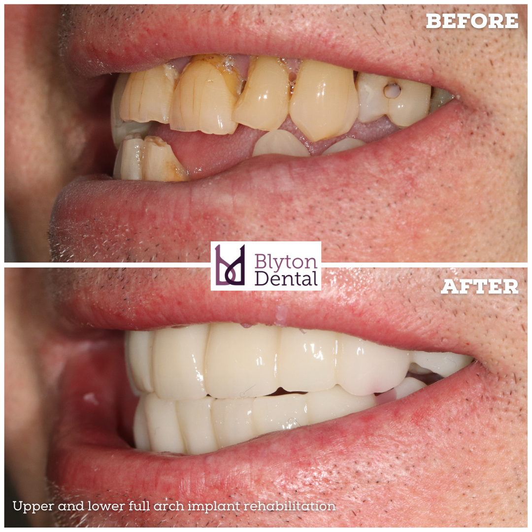
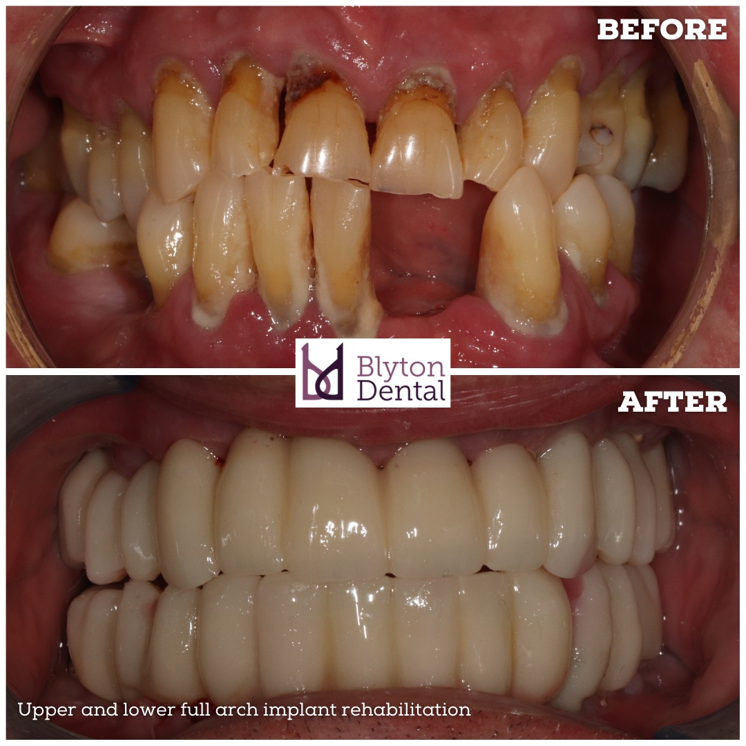
This gentleman came to us with a poor state of the remaining dentition.
Severe gum disease and and anterior cross bite made the remaining teeth unrestorable.
A full digital protocol was adopted using clinical photographs, 3D impressions with trios 5 scanner and CBCT scans.
3 shape software was used to design his diagnostic mock ups and implant guides.
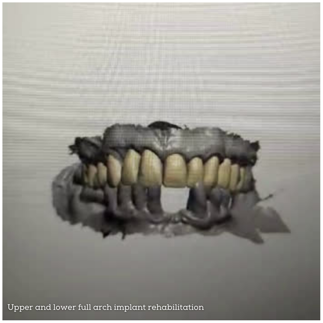
All the remaining teeth were extracted, six implants were placed in each jaws immediately followed by digital impressions with trios 5, and immediate implant bridges (milled PMMA) were delivered.
He was very happy with results - these post op images are just 3 days after surgery.
If everything goes well, he will receive his long term definitive zirconia bridges in 3 months.
Over the last two years or so, we have transitioned to this fully digital protocol from a conventional chair side conversion. This is definitely the way forward in FP1 and most FP2 scenarios.